
Why Standardized Measurements Matter
Hoof radiographs contain a wealth of information about the internal structures of the foot. But that information is only useful when it’s presented in a clear, repeatable way that both the veterinarian and the farrier can rely on.
Standardized measurements transform subjective visual impressions into objective data points — making it possible to compare findings across visits, track changes over time, and communicate precisely about what needs attention.
Without consistent measurement methodology, two radiographs of the same foot taken weeks apart can tell very different stories depending on positioning, interpretation approach, and reporting format.
Key Measurements in Hoof Radiograph Reports
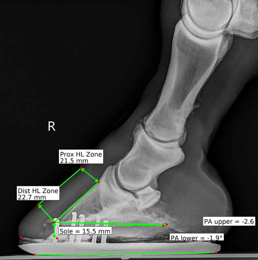
Sole Depth
Sole depth is one of the most commonly referenced measurements in hoof radiograph reports. It represents the distance from the distal border of the third phalanx (P3) to the ground surface of the sole.
Monitoring sole depth over time helps veterinarians and farriers:
- Assess whether the horse has adequate protection
- Identify thinning patterns that may indicate pathology
- Guide pad and shoe selection decisions
- Track recovery progress in cases of laminitis or sole bruising
Palmar Angle
The palmar angle describes the angle of the solar surface of P3 relative to the ground. This measurement is particularly significant in laminitis cases and when evaluating hoof balance.
A negative palmar angle may indicate:
- Distal displacement of the coffin bone
- Chronic laminitic changes
- The need for therapeutic shoeing intervention
Hoof-Pastern Axis
The hoof-pastern axis (HPA) evaluates the alignment of the phalanges from the proximal phalanx through the middle phalanx to the distal phalanx. A broken-back or broken-forward axis can indicate hoof balance issues that affect the entire distal limb.
Mediolateral Balance
Lateromedial radiographic views can reveal asymmetric loading patterns. Measuring the relative heights of the medial and lateral joint spaces helps assess whether the foot is bearing weight symmetrically.
How DigiHoof Standardizes These Measurements
DigiHoof uses consistent landmark placement and automated measurement calculation to ensure that every report follows the same methodology. This means:
- Repeatable results — the same landmarks are identified and measured the same way every time
- Visual overlays — measurements are displayed directly on the radiograph image
- Clear formatting — values are presented in a structured report layout that’s easy to read and reference
AI-Assisted Landmark Detection
DigiHoof’s machine learning system assists with detecting key anatomical landmarks on the radiograph. This helps normalize landmark placement across cases while preserving the veterinarian’s ability to verify and adjust any point before the final report is generated.
The goal of AI assistance is not to replace clinical judgment but to make the reporting process fast enough to be practical in high-volume imaging days.
Using Measurements to Guide Farrier Decisions
When a farrier receives a DigiHoof report, they see the same standardized measurements and overlays the veterinarian reviewed. This shared visual language enables more productive conversations about:
- How much sole depth is available before aggressive trimming
- Whether the palmar angle needs correction
- What type of shoe or pad configuration might benefit the horse
- How the foot has changed since the last evaluation
Longitudinal Tracking
One of the most powerful aspects of standardized measurements is the ability to compare across visits. When every report uses the same methodology, real trends emerge:
- Sole depth progression over a trimming cycle
- Palmar angle response to therapeutic shoeing
- Hoof-pastern axis improvement with corrective trimming
- Mediolateral balance changes over time
Best Practices for Radiograph Quality
The accuracy of any measurement depends on the quality of the original radiograph. For the best results:
- Positioning — ensure consistent foot placement and beam angle
- Markers — use a radiopaque dorsal wall marker when possible
- Labeling — clearly identify left vs. right, and lateral vs. dorsopalmar views
- Consistency — try to replicate the same setup across follow-up visits
Conclusion
Standardized hoof radiograph measurements are the foundation of effective veterinary-farrier communication. By ensuring that every report speaks the same language, DigiHoof helps both professionals focus on what matters most — making the best decisions for the horse.